A Microscopic Look at Snail Jaws
Have you ever wondered what the inside of a snail's mouth looks like?

Have you ever wondered what the inside of a snail's mouth looks like?
The anatomy involved in land snail and slug feeding is fascinating. Well, I’d like to guess that it is more fascinating than you’d expect if you’ve ever thought about snail and slug feeding in the first place. Snails and slugs have evolved to eat just about everything; they are herbivorous, carnivorous, omnivorous, and detritivorous (eating decaying waste from plants and other animals). There are specialist and generalist species that eat worms, vegetation, rotting vegetation, animal waste, fungus, and other snails.
Thousands of Microscopic Teeth!
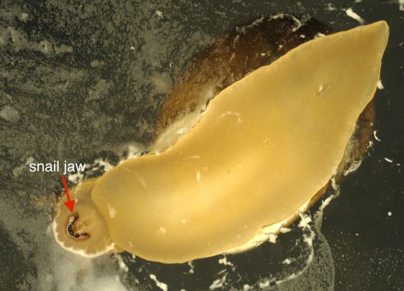
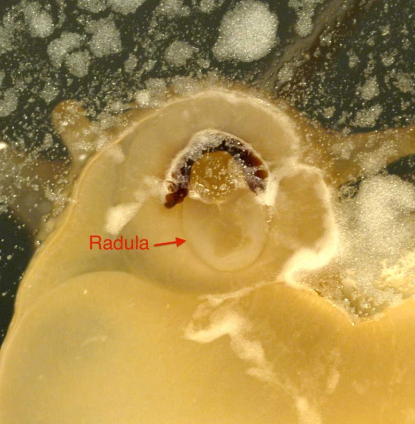
Snails and slugs eat with a jaw and a flexible band of thousands of microscopic teeth, called a radula. The radula scrapes up, or rasps, food particles, and the jaw cuts off larger pieces of food, like a leaf, to be rasped by the radula. To understand what the single jaw and radular band look like in a terrestrial snail, two Museum interns (from Glendale Community College), Ala Babakhanians and Richard Laguna, photographed a common European Garden Snail (Cornu aspersum) eating a film of cornstarch and water on a piece of glass. This clever method was inspired by the Snail's Tales blog.
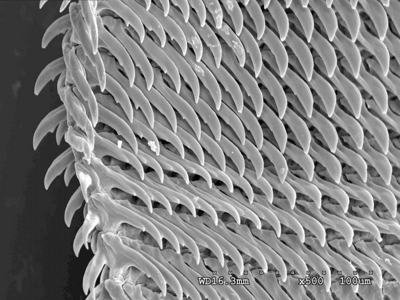
A Close Look at a Slug's Rasping Radula
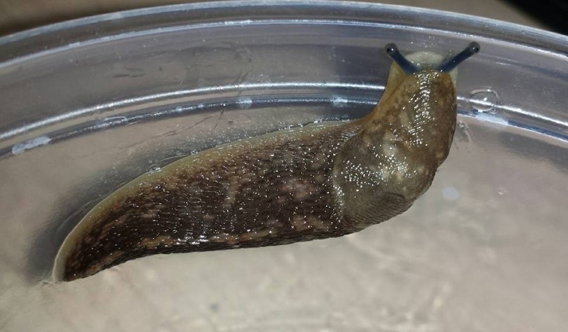
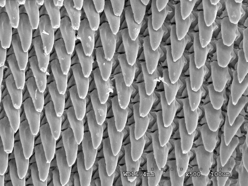
The only way to truly appreciate the microscopic teeth of the radula is to look at them under a microscope. To do this, Ole Willadsen, another Glendale Community College intern at NHMLA, dissected out the radula from a non-native slug found on Sunset Boulevard.
The radula was imaged using the Museum's scanning electron microscope (SEM), which creates an extremely detailed and highly magnified picture of the specimen examined.
The fascinating feeding anatomy of snails and slugs is also helpful in determining their species identity, if and when that is in question. Since we sometimes don’t know the identity of non-native species we encounter in Los Angeles, the size and shape of their single jaw and thousands of radular teeth can be as informative as they are beautiful.
**Can you hear a snail eating? Yes! Check out Elisabeth Tova Bailey’s book The Sound of a Wild Snail Eating.
If you’d like to be involved in efforts to document and protect L.A.’s biodiversity, check out our Community Science program. Or you can donate to the Urban Nature Research Center.
(Posted by: Jann Vendetti)

